Facial Trauma · Volume I of 6
Seeing the Injured Face
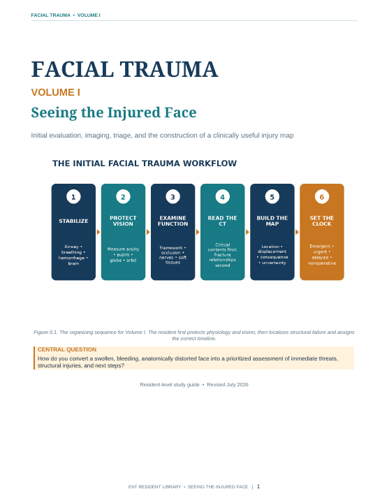
Turn a swollen or bleeding face into a time-sensitive injury map by protecting airway and vision, performing a reproducible examination, reviewing CT systematically, and assigning urgency.
Orientation
About this volume
Volume I establishes the assessment framework for the Facial Trauma series before regional repair begins. It organizes the first consultation around physiology, vision, the structural logic of the face, a functional examination, and an eight-pass CT review. Residents learn to separate fracture names from threatened function; identify airway, hemorrhagic, ocular, neurologic, and skull-base concerns; document limitations; and build a concise injury map with the right timeline. Integrated trauma-bay cases reinforce presentation and triage. Unlike the following regional volumes, this opening volume deliberately stops before detailed exposure, reduction, fixation, and reconstruction. It prepares residents to recognize what is immediately dangerous, what requires urgent specialist input, and what can be planned safely.
After completing this volume, the resident should be able to
- Prioritize airway, hemorrhagic, ocular, neurologic, and skull-base threats before fracture repair.
- Perform and document a reproducible facial examination and independent multi-plane CT review.
- Present a functional injury map with emergent, urgent, delayed-operative, or nonoperative priorities.
Reader
PDF preview
Preview before loading
The full PDF is loaded only when requested to keep this page fast, especially on mobile.