Facial Trauma · Volume II of 6
Orbit, Frontal Sinus, and Anterior Skull Base
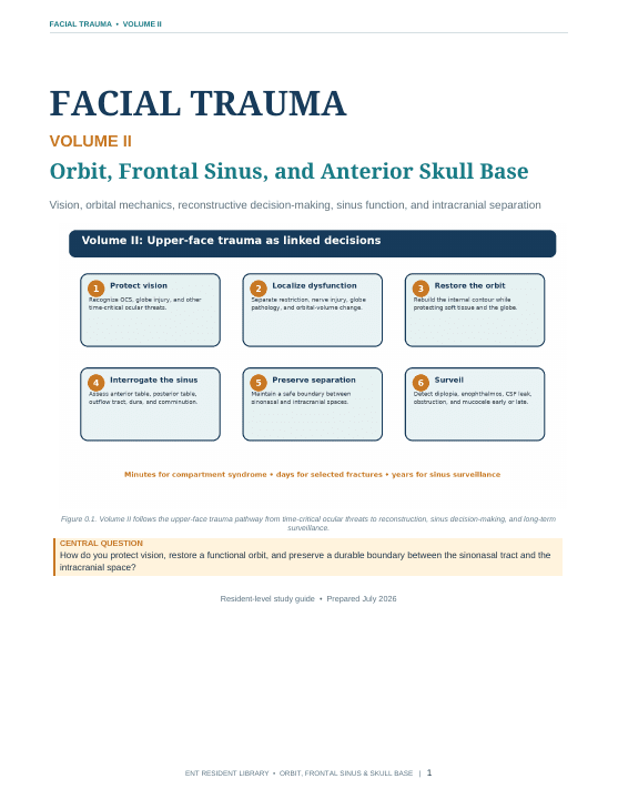
Manage upper-face trauma through vision, orbital mechanics, sinus drainage, and intracranial separation—localizing diplopia, planning reconstruction, and applying a structured frontal-sinus decision framework.
Orientation
About this volume
Volume II narrows the initial trauma framework to the upper face, where orbit, frontal sinus, and anterior skull base injuries share thin bony boundaries and competing functional priorities. Residents move from the threatened eye and orbital fracture evaluation to exposure, implant geometry, and postoperative checks, then upward to frontal-sinus drainage, posterior-table injury, CSF leak, and surveillance. The guide’s five-variable frontal-sinus worksheet links anatomy and reliability of follow-up to preservation, contour repair, obliteration, or cranialization decisions. It assumes the trauma survey and complete examination from Volume I. Unlike Volume III’s central and lateral midface reconstruction, this volume focuses on protecting vision and maintaining durable separation between sinonasal and intracranial spaces.
After completing this volume, the resident should be able to
- Localize orbital trauma through vision, motility, globe position, and CT findings while recognizing time-critical ocular threats.
- Plan orbital reconstruction around defect geometry, exposure, implant verification, and postoperative surveillance.
- Use anterior-table, posterior-table, outflow-tract, dural, and comminution findings to frame frontal-sinus decisions.
Reader
PDF preview
Preview before loading
The full PDF is loaded only when requested to keep this page fast, especially on mobile.